| Q. |
What are bowlegs? |
 |
| A. |
Bowleg is a condition in which the legs have a gradual or sudden bent due to various reasons. In medical terms we call them varus (bent inwards) or valgus (bent outward). |
|
| |
|
|
| Q. |
How does that affect me? |
|
| A. |
Normally, on standing both the knees bear equal weight, and within the knee itself the load is borne equally by the inner and outer side. When a person has bowlegs there is increased load on the inner side of the knee more commonly (Varus) or the outer side of the knee (Valgus). This increased loading causes that side to wear out faster and then presents as pain. |
|
| |
|
|
| Q. |
But I have the bow since childhood, then why is it worsening now? |
|
| A. |
Generally the body is able to compensate slight bow in the legs by the action of muscles. But when the bent is beyond a certain limit then the muscles cannot compensate and this results in reduction in the joint space and gradual worsening of the angle. |
|
| |
|
|
| Q. |
What do you mean by joint space? |
|
| A. |
The junction of two bones for the purpose of smooth movement makes the joint. To help in the
smooth gliding motion of the bones, the bone ends have a covering called cartilage. This cartilage
is actually a very smooth, shiny and delicate structure. Only the bone is visible on the x-ray
because of its calcium content. The cartilage is not seen on x-ray and there appears to be a gap in
the joint, which is referred to as joint space. |
|
| |
|
|
| Q. |
WhI have been diagnosed as reduced joint space/uni-compartmental arthritis/varus deformity.
What is the meaning of this? |
|
| A. |
When the cartilage of the joint wears out it is called reduced joint space. As this becomes more advanced it starts affecting the underlying bone also and its then called arthritis. Unicompartmental
means only one of the three compartments of the knee are affected. |
|
| |
|
|
| Q. |
Do all bowlegs need surgery? |
|
| A. |
No, we advise surgery only when there is significant pain along with the deformity. |
|
| |
|
|
| Q. |
What is significant pain? |
|
| A. |
When there is enough pain on walking or standing that does not settle with exercises and requires daily pain medication. |
|
| |
|
|
| Q. |
Doctor, I have only occasional pain, which settles with rest. I am worried that this will
worsen and I’ll be crippled. What should I do? |
|
| A. |
Occasional pain that settles with rest doesn’t require surgery immediately. Generally in about 80-90% people this settles down with proper exercises and medication for a few days. The only way then to reap the benefits is to continue exercises religiously daily. |
|
| |
|
|
| Q. |
Does this mean I’ll be able to avoid the surgery all together? |
|
| A. |
Yes and no.
We keep you under follow up. If there is no worsening on the x-rays (done yearly) and there is no pain with exercises then nothing more than a yearly follow-up is required. But if there is worsening on the x-rays even without much pain, then there is a role of correction of the deformity as a “preventive surgery”. |
|
| |
|
|
| Q. |
What are the surgical choices? |
|
| A. |
The choices are HTO, Knee Replacement, and Arthroscopy. |
|
| |
|
|
| Q. |
What is HTO? |
|
| A. |
HTO is for High Tibial Osteotomy – high means up or close to knee joint or proximal, Tibial is for the leg bone Tibia below the knee joint, and Osteotomy is for cutting the bone. |
|
| |
|
|
| Q. |
Why is it required? |
|
| A. |
The bone is cut to change the angle of the bone and correct the bow. |
|
| |
|
|
| Q. |
How does the change of angle help? |
|
| A. |
By changing the angle of bone the bow is corrected and the load-bearing axis of the bone is brought back to the centre of the knee joint. This shift helps in taking the load off the affected side and relieves the pain, but more importantly it helps the overloaded side to get rest and heal. As the healing progresses the narrowed joint space again fills up cartilage. |
|
| |
|
|
| Q. |
You mean the cartilage grows back! Doctor I had heard that once the cartilage is damaged, it never comes back, is it true? |
|
| A. |
It is true that the cartilage (Hyaline cartilage), which normally covers the joint surface of the bone never, comes back, because it does not have the capacity to regenerate. But when it is given adequate rest and opportunity to heal it heals with a different kind of cartilage structurally (Fibro-cartilage) that is functionally similar to the original cartilage. |
|
| |
|
|
| Q. |
That sounds interesting, could you elaborate on that a little more? |
|
| A. |
Certainly, let me explain with an example – everybody knows about a car tyre. It has a metallic rim covered with a tube and tyre. The rim is the actual thing driven by the motors. To protect the rim and also to reduce the friction with the road the tube and tyre provide an outer coating. If the rim is not aligned properly then the tyre wears out from one side faster. So to correct the problem of a worn out tyre one can either put a new tyre or correct the alignment. If just a new tyre is put without correcting the alignment then the problem would be solved temporarily but the new tyre would also wear out in sometime, whereas if the wheel balancing (alignment correction) is done then the wear reduces to the normal rate.
Similarly in the human body the rim is the bone and the protective tyre is the cartilage. When there is a problem of alignment of the bones as in bowlegs the cartilage of one side wears out faster. As the loading is constant with activity the cartilage doesn’t get a chance to heal and the damage continues to progress ultimately causing the bones to rub on each other. Here again the same options hold true – replacement or realignment. Replacement without realignment will cause wear of the new joint. Realignment will unload the affected side and give a chance for natural healing to occur. |
|
| |
|
|
| Q. |
Is there any special test required before HTO? |
|
| A. |
Every patient requires certain x-rays: standing x-rays of both knees and both legs (Full Length x-rays) besides lateral x-ray of the affected side in maximum extension.
Occasionally MRI of the knee may also be required to document the amount of cartilage damage and bone marrow edema.
A list of blood investigations is also performed to check pre-operative fitness. |
|
| |
|
|
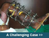
| Q. |
How is the HTO performed? |
|
| A. |
The principle is to cut the Tibia through a small incision (about 1.5 cm) few inches below the knee; an external fixator is applied to the bone; the angle is then gradually corrected by turning a nut on the fixator (1 mm per day) to a pre-defined point and the fixator is then locked. |
|
| |
|
|
| Q. |
Does all of this is get done during the surgery? |
|
| A. |
The fixator is applied and the bone is cut during the surgery, so when you are out of the operation theatre your leg is still bent. After 7 days the distraction is started and with that the angle is gradually corrected over a period of about 3 weeks, depending on the amount of correction required. |
|
| |
|
|
| Q. |
How long does the surgery last? |
|
| A. |
The surgery itself is about 45-60 min but the anesthesia and recovery etc add up to make it 1.5 to 2 hours. |
|
| |
|
|
| Q. |
What kind of anesthesia is used? |
|
| A. |
The anesthetist is the best judge for the type of anesthesia, but generally we prefer spinal or epidural anesthesia. |
|
| |
|
|
| Q. |
How soon do I recover from the surgery? |
|
| A. |
You are conscious at the end of the procedure, though you may feel a little sleepy for a short while. |
|
| |
|
|
| Q. |
Is the procedure painful? |
|
| A. |
The minimally invasive technique we use for the surgery and the other techniques to minimize any soft tissue damage helps to keep the pain to a minimum. We also take other adequate measures to keep the patient comfortable. |
|
| |
|
|
| Q. |
How long do I have to take bed rest? |
|
| A. |
By evening you are taking diet by mouth. We advise rest for 24 hours, after which gradual sitting in bed is allowed and mobilization is begun. Even while in bed it is not a complete rest rather we encourage gentle ankle and toe movements and knee exercises. |
|
| |
|
|
| Q. |
When do I start walking? |
|
| A. |
After the second day walking is allowed using a walker. First you start walking touch down weight bearing, i.e. just touching the foot down without really taking weight on the leg (about 5 kg is allowed – we say “imagine putting an egg or a tomato under your foot, and you don’t crush it”).
Once the fixator is locked weight bearing is gradually increased, the walker is then switched to cane/stick so that towards the end of the fixator period you are walking full weight bearing without any support. |
|
| |
|
|
| Q. |
How long do I have to stay in the hospital? |
|
| A. |
Hospital stay is 5-7 days. From the point of view of the surgery and small incisions discharge can be given in 2 days but we keep you for a longer time to teach you the importance of exercises, the way to take care of the fixator and the way to distract. |
|
| |
|
|
| Q. |
How many times do I have to come for follow-up? |
|
| A. |
After discharge the first visit is for suture removal (generally only 2 or 3 sutures) at the end of 2 weeks from surgery. Thereafter a visit is required every 10 days or so till the fixator is locked. Once the fixator is locked then a visit is required once in 3-4 weeks. On every visit x-ray is done.
A full-length x-ray is done at the end of distraction, to determine the complete correction. After removal of the fixator another visit is recommended after 2 weeks and thereafter every 6 months to a year. |
|
| |
|
|
| Q. |
Doctor, what is this distraction? |
|
| A. |
Distraction is the process by which the cut ends of the bone are pulled apart. When it is done at a certain rate gradually, new bone forms between the bone ends. This is the advantage of the Ilizarov method of treatment. |
|
| |
|
|
| Q. |
Is this new bone any different from the original bone? |
|
| A. |
No, once the new bone matures or is strong enough in 6-8 weeks, it is just like the original bone. So one year down the line nobody can even make out where the bone was cut. |
|
| |
|
|
| Q. |
How is the fixator locked? Is it painful? |
|
| A. |
Turning a nut on the fixator locks the fixator. But before that a full-length x-ray is done, the angles are measured and only when they are adequate, the fixator is locked. We use very stringent measures to define the correction, as research has shown the maximum longevity of the knee when the correction is done within a narrow range of 3 degrees. So, if we find there is still some more correction required then that many days of correction is advised and only then the fixator is locked, after a repeat full-length x-ray. No, it is not painful at all. The process is done on OPD basis. |
|
| |
|
|
| Q. |
How long does the fixator remain? |
|
| A. |
The fixator remains for a total of 3 months, because that is the time for the bone to heal and become solid. |
|
| |
|
|
| Q. |
But I’m from out of station; do I need to stay in Mumbai for the complete treatment? |
|
| A. |
Not necessarily. We operate on many patients from out of town. These patients are discharged at the end of 7 days, only when they have understood the Pin care site, exercises and the distraction process and we are satisfied with their progress.
The sutures can be taken out at your hometown. The x-rays are prescribed on the discharge note and can be sent either by courier or by email.
At the end of distraction (the days are pre-calculated and informed accordingly), a re-visit to Mumbai needs to be planned for a day. During this visit a full-length x-ray is done and according to the Correction achieved any changes if required, are made and the fixator is locked. This is an OPD procedure and you can travel back later on the same day.
Thereafter x-rays are to be sent via courier/email once every 3-4 weeks. At the end of 3 months, the fixator is removed, which is again an OPD procedure, and travel can be planned later on the same day. |
|
| |
|
|
| Q. |
Does fixator removal require surgery? |
|
| A. |
No, the fixator is removed in the OPD itself. |
|
| |
|
|
| Q. |
How soon can I start normal activities after the surgery? |
|
| A. |
The patient is usually independent and doesn’t require anybody to take care of him/her. Generally office going can be started by 4 weeks i.e. after the fixator is locked. Though there is no restriction for resuming work, some of the patients find it difficult to manage going to the office with the walker.
Driving is not recommended till 8-10 weeks post surgery.
The housewives require help for the chores, though they are able to supervise the work. |
|
| |
|
|
| Q. |
Are there any precautions to be taken after removal of fixator? |
|
| A. |
A cane/stick should be used for 2 weeks after the removal of fixator. This is more of a precaution to remind the patient to take things light and also for other people to let them know and avoid any pushing or shoving. |
|
| |
|
|
| Q. |
How long does it take to appreciate the benefits of the surgery? |
|
| A. |
Almost immediately. The knee pain which you had before the surgery is immediately releived. The post surgery pain is minimum, which settles down with anti-inflammatory medication within a couple of days.
However, to continue with the pain relief for a long term, a simple and regular exercise program needs to be followed.
The joint space (or the cartilage) recovery to show up on the x-ray takes about an year. |
|
| |
|
|
| Q. |
Do I need to modify my lifestyle to protect my knees from further damage? |
|
| A. |
Not really!
Whatever you were able to do before surgery, you would be still able to do, if at all the activities would be easier to perform. On the long term basis activities like swimming, walking, cycling without tension on the belt, treadmill without too much incline are all good, whereas jogging especially on hard surfaces like cemented tracks, cycling with tension are preferably to be avoided as they put undue stress and load on the knee joint We recommend a "common sense" approach, to avoid or minimize the activites which cause pain or difficulty. In the post surgical period once you are back to the pre-surgery status, gradual resumption and addition of activites should be done. |
|
| |
|
|